

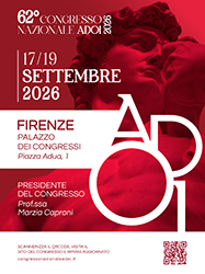
62° Congresso Nazionale ADOI, Firenze, 17-19 settembre 2026
Vol. 18 No. s1 (2026): 62° Congresso Nazionale ADOI, Firenze, 17-19 settembre 2026

14 | CUTANEOUS LEISHMANIASIS MIMICKING B-CELL PSEUDOLYMPHOMA: A DIAGNOSTIC CHALLENGE IN AN ENDEMIC MEDITERRANEAN AREA
M. Fino1, A. Zordan1, N. Di Meo1, I. Zalaudek1, S. Sola2, C. Massone3 | 1Department of Dermatology and Venereology, University of Trieste, Maggiore Hospital, Trieste, Italy; 2Department of Pathology, Galliera Hospital, Genoa, Italy; 3Department of Dermatology, Galliera Hospital, Genoa, Italy
Publisher's note
All claims expressed in this article are solely those of the authors and do not necessarily represent those of their affiliated organizations, or those of the publisher, the editors and the reviewers. Any product that may be evaluated in this article or claim that may be made by its manufacturer is not guaranteed or endorsed by the publisher.
All claims expressed in this article are solely those of the authors and do not necessarily represent those of their affiliated organizations, or those of the publisher, the editors and the reviewers. Any product that may be evaluated in this article or claim that may be made by its manufacturer is not guaranteed or endorsed by the publisher.
Published: 24 September 2026
0
Views
0
Downloads
Authors
Downloads
Download data is not yet available.
Citations



How to Cite
1.
14 | CUTANEOUS LEISHMANIASIS MIMICKING B-CELL PSEUDOLYMPHOMA: A DIAGNOSTIC CHALLENGE IN AN ENDEMIC MEDITERRANEAN AREA: M. Fino1, A. Zordan1, N. Di Meo1, I. Zalaudek1, S. Sola2, C. Massone3 | 1Department of Dermatology and Venereology, University of Trieste, Maggiore Hospital, Trieste, Italy; 2Department of Pathology, Galliera Hospital, Genoa, Italy; 3Department of Dermatology, Galliera Hospital, Genoa, Italy. Dermatol Reports [Internet]. 2026 Sep. 24 [cited 2026 Sep. 24];18(s1). Available from: https://journals.pagepress.net/dr/article/view/11081
Copyright (c) 2026 The Author(s)


This work is licensed under a Creative Commons Attribution-NonCommercial 4.0 International License.
