


62° Congresso Nazionale ADOI, Firenze, 17-19 settembre 2026
Vol. 18 No. s1 (2026): 62° Congresso Nazionale ADOI, Firenze, 17-19 settembre 2026

33 | GENITAL POROKERATOSIS CONFIRMED BY HISTOPATHOLOGY AND REFLECTANCE CONFOCAL MICROSCOPY: A RARE CASE REPORT
G. Frattin1|2, A. Bolzon1|2, E. Pezzolo1, L. Germi1, L. Gnesotto1, A. Corrà1, G. Mioso1, Z. Fratton1, L. Schiesari1 | 1UOC Dermatologia, Ospedale San Bortolo, Vicenza, Italy; 2Università degli Studi di Padova, Clinica Dermatologica, Italy
Publisher's note
All claims expressed in this article are solely those of the authors and do not necessarily represent those of their affiliated organizations, or those of the publisher, the editors and the reviewers. Any product that may be evaluated in this article or claim that may be made by its manufacturer is not guaranteed or endorsed by the publisher.
All claims expressed in this article are solely those of the authors and do not necessarily represent those of their affiliated organizations, or those of the publisher, the editors and the reviewers. Any product that may be evaluated in this article or claim that may be made by its manufacturer is not guaranteed or endorsed by the publisher.
Published: 24 September 2026
0
Views
0
Downloads
Authors
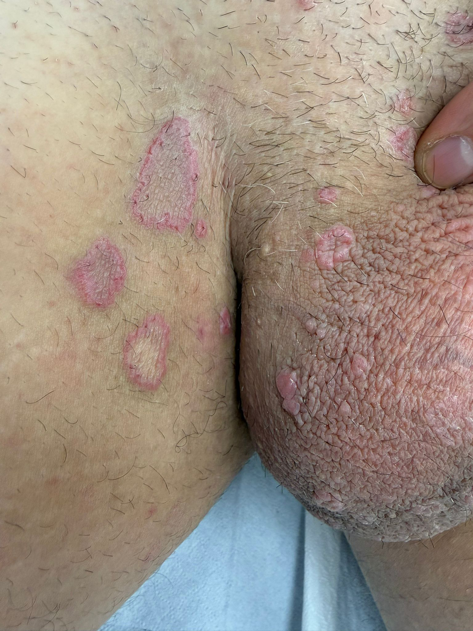
Materials and Methods. A 45-year-old otherwise healthy man, with no history of immunosuppression or sexually transmitted infections, presented with multiple pruritic annular plaques involving the inguinal region, inner thigh, penile base and scrotum. The lesions had been present for approximately four years. Clinical examination, RCM and histopathological assessment were performed. A biopsy was obtained from the raised keratotic border of a representative lesion on the right inner thigh.
Results. RCM showed a well-defined peripheral rim corresponding to the clinically visible keratotic border, associated with focal interruption of the epidermal architecture and loss of the physiological honeycomb pattern. Hyperreflective parakeratotic material within the epidermal furrow was observed, suggesting the in vivo counterpart of cornoid lamellae. Histopathology revealed compact hyperkeratosis, focal erosion, moderate-to-severe irregular acanthosis, moderate spongiosis with mild lymphocytic exocytosis, and two areas of dyskeratosis surmounted by marked hyperparakeratosis, consistent with cornoid lamellae. A moderate discontinuous band-like lymphocytic infiltrate was present in the superficial dermis. PAS, PASD and Alcian blue stains were negative for fungal elements; immunohistochemistry for herpes simplex virus and Treponema pallidum was negative. Previous treatment with topical corticosteroids had been ineffective, and the patient is currently receiving acitretin 25 mg/day with clinical follow-up.
Conclusions. This case highlights genital porokeratosis as a rare and underrecognized diagnosis in chronic annular genital and paragenital plaques. The combined use of clinical examination, RCM and targeted histopathology may improve diagnostic accuracy, support biopsy site selection and strengthen clinicopathological correlation in anatomically sensitive areas.
Downloads
Download data is not yet available.
Citations



1. Valdivielso-Ramos M. Genital porokeratosis. Actas Dermosifiliogr. 2008;99:217-220.
2. Dongre A, Adhe V, Sanghavi S. Genital porokeratosis: a rare entity. Indian Journal of Dermatology. 2013;58:81-83.
3. Pietkiewicz P, Korecka K, Salwowska N, Kohut I, Adhikari A, Bowszyc-Dmochowska M, Pogorzelska-Antkowiak A, Navarrete-Dechent C. Porokeratoses - A comprehensive review on the genetics and metabolomics, imaging methods and management of common clinical variants. Metabolites. 2023;13:1176.
How to Cite
1.
33 | GENITAL POROKERATOSIS CONFIRMED BY HISTOPATHOLOGY AND REFLECTANCE CONFOCAL MICROSCOPY: A RARE CASE REPORT: G. Frattin1|2, A. Bolzon1|2, E. Pezzolo1, L. Germi1, L. Gnesotto1, A. Corrà1, G. Mioso1, Z. Fratton1, L. Schiesari1 | 1UOC Dermatologia, Ospedale San Bortolo, Vicenza, Italy; 2Università degli Studi di Padova, Clinica Dermatologica, Italy. Dermatol Reports [Internet]. 2026 Sep. 24 [cited 2026 Sep. 24];18(s1). Available from: https://journals.pagepress.net/dr/article/view/11094
Copyright (c) 2026 The Author(s)


This work is licensed under a Creative Commons Attribution-NonCommercial 4.0 International License.
