


62° Congresso Nazionale ADOI, Firenze, 17-19 settembre 2026
Vol. 18 No. s1 (2026): 62° Congresso Nazionale ADOI, Firenze, 17-19 settembre 2026

40 | BEYOND THE USUAL CAT: TRICHOPHYTON BENHAMIAE KERION CELSI IN A CHILD WITH CHINCHILLA-ASSOCIATED HOUSEHOLD OUTBREAK
T. Galeotti1, D. Tesauri1, P. Caposciutti1, L. Bianchi1, S. Crotti2, D. Cruciani2, K. Hansel1, L. Stingeni1 | 1Dermatology Unit, Department of Medicine and Surgery, University of Perugia, Perugia, Italy; 2Istituto Zooprofilattico Sperimentale dell'Umbria e delle Marche "Togo Rosati", Perugia, Italy
Publisher's note
All claims expressed in this article are solely those of the authors and do not necessarily represent those of their affiliated organizations, or those of the publisher, the editors and the reviewers. Any product that may be evaluated in this article or claim that may be made by its manufacturer is not guaranteed or endorsed by the publisher.
All claims expressed in this article are solely those of the authors and do not necessarily represent those of their affiliated organizations, or those of the publisher, the editors and the reviewers. Any product that may be evaluated in this article or claim that may be made by its manufacturer is not guaranteed or endorsed by the publisher.
Published: 24 September 2026
0
Views
0
Downloads
Authors
Materials and Methods. We report a pediatric case of extensive dermatophytosis initially managed in the outpatient setting and subsequently hospitalized due to clinical worsening. Clinical evaluation, laboratory testing, mycological examination, and epidemiological reconstruction were performed. Definitive species identification was obtained through molecular sequencing.
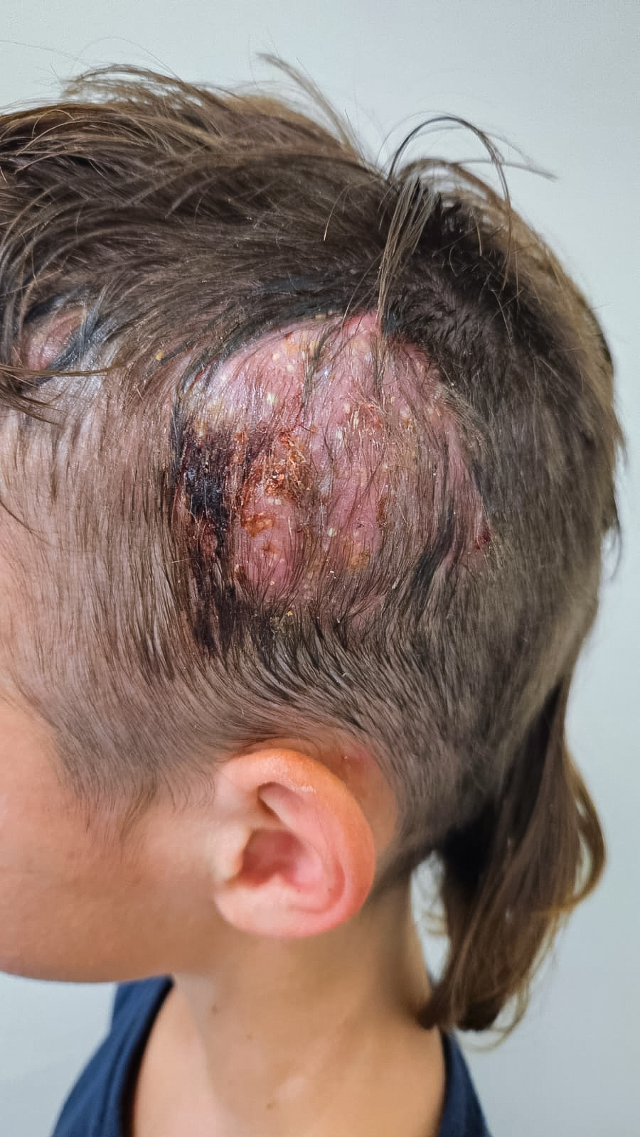
Results. A 5-year-old boy presented with multiple erythematous, scaly papulo-squamous lesions involving the face, trunk, gluteal region, and scalp. Initial outpatient management by a community dermatologist included topical antifungal therapy for suspected tinea corporis, with partial improvement. Over the following weeks, scalp involvement progressed to a large inflammatory alopecic plaque with pustulation, crusting, and associated retroauricular and laterocervical lymphadenopathy, consistent with kerion celsi. The patient was therefore referred for hospital admission. On admission, laboratory tests showed mild neutrophilic leukocytosis and elevated inflammatory markers. Systemic therapy with griseofulvin was initiated, together with ampicillin/sulbactam for suspected bacterial superinfection and topical antiseptic/antibacterial treatment (fusidic acid and aqueous fuchsin). Bacterial and yeast cultures from skin swabs were negative. During hospitalization and subsequent follow-up visits, progressive clinical improvement was observed, with crusting of lesions, reduction of the pustular component, and normalization of inflammatory markers. Species identification as Trichophyton benhamiae was achieved by fungal culture and confirmed by molecular sequencing. Epidemiological re-evaluation revealed a household zoonotic outbreak. A sibling presented with concomitant tinea corporis of the trunk, successfully treated with topical terbinafine. Domestic chinchillas showed alopecia and pruritus and were subsequently treated, strongly suggesting an animal reservoir as the source of infection. Initial exposure hypotheses, including animal contact during a travel in India and rural accommodation, were considered less relevant. At follow-up, the patient showed marked clinical improvement under systemic antifungal therapy, with resolution of inflammatory signs and residual patchy alopecia of the scalp.
Conclusions. This case highlights a household outbreak of Trichophyton benhamiae infection presenting with extensive tinea corporis and kerion celsi in a child, confirmed by molecular identification. It emphasizes the importance of repeated and dynamic epidemiological assessment and awareness of atypical zoonotic reservoirs, such as chinchillas and guinea pigs. Molecular diagnostics can be crucial for confirming emerging dermatophyte species and for accurate outbreak tracing.
Downloads
Download data is not yet available.
Citations



1. Nenoff P, Uhrlass S, Kruger C, et al. Trichophyton species of Arthroderma benhamiae - a new infectious agent in dermatology. J Dtsch Dermatol Ges. 2014;12:571-581.
2. Nenoff P, Schulze I, Uhrlass S, Kruger C. Kerion caused by the zoophilic dermatophyte Trichophyton species of Arthroderma benhamiae in a child. Hautarzt. 2013;64:846-849.
3. Hattori N, Kaneko T, Tamaki K, Makimura K, Mochizuki T. A case of kerion celsi due to Arthroderma benhamiae identified by DNA sequences of nuclear ribosomal internal transcribed spacer 1 regions. Med Mycol. 2003;41:249-251.
4. Shiraki Y, Hiruma M, Kano R, Miyamoto C, Ikeda S. Case of tinea capitis caused by Trichophyton mentagrophytes (molecular type Arthroderma benhamiae). J Dermatol. 2006;33:504-506.
5. Bartosch T, Frank A, Gunther C, et al. Trichophyton benhamiae and T. mentagrophytes target guinea pigs in a mixed small animal stock. Med Mycol Case Rep. 2018;23:37-42.
How to Cite
1.
40 | BEYOND THE USUAL CAT: TRICHOPHYTON BENHAMIAE KERION CELSI IN A CHILD WITH CHINCHILLA-ASSOCIATED HOUSEHOLD OUTBREAK: T. Galeotti1, D. Tesauri1, P. Caposciutti1, L. Bianchi1, S. Crotti2, D. Cruciani2, K. Hansel1, L. Stingeni1 | 1Dermatology Unit, Department of Medicine and Surgery, University of Perugia, Perugia, Italy; 2Istituto Zooprofilattico Sperimentale dell’Umbria e delle Marche "Togo Rosati", Perugia, Italy. Dermatol Reports [Internet]. 2026 Sep. 24 [cited 2026 Sep. 24];18(s1). Available from: https://journals.pagepress.net/dr/article/view/11100
Copyright (c) 2026 The Author(s)


This work is licensed under a Creative Commons Attribution-NonCommercial 4.0 International License.
