


62° Congresso Nazionale ADOI, Firenze, 17-19 settembre 2026
Vol. 18 No. s1 (2026): 62° Congresso Nazionale ADOI, Firenze, 17-19 settembre 2026

57 | GENERALIZED PLANE XANTHOMA AND NECROBIOTIC XANTOGRANULOMA HERALDING PROGRESSION FROM MGUS TO MULTIPLE MYELOMA: COMPLETE CUTANEOUS RESOLUTION AFTER MYELOMA-DIRECTED THERAPY
M. Di Prete1, V. Lora2, C. Cota1 | 1Dermatopathology Research Unit, San Gallicano Dermatological Institute IRCCS, Rome, Italy; 2Clinical Dermatology, San Gallicano Dermatological Institute IRCCS, Rome, Italy
Publisher's note
All claims expressed in this article are solely those of the authors and do not necessarily represent those of their affiliated organizations, or those of the publisher, the editors and the reviewers. Any product that may be evaluated in this article or claim that may be made by its manufacturer is not guaranteed or endorsed by the publisher.
All claims expressed in this article are solely those of the authors and do not necessarily represent those of their affiliated organizations, or those of the publisher, the editors and the reviewers. Any product that may be evaluated in this article or claim that may be made by its manufacturer is not guaranteed or endorsed by the publisher.
Published: 24 September 2026
0
Views
0
Downloads
Authors
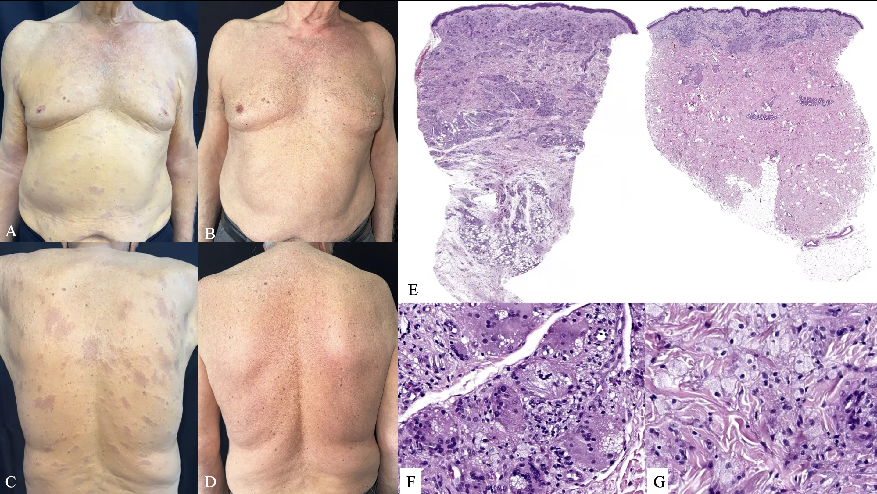
Materials and Methods. An 80-year-old man with a 17-year history of IgG lambda MGUS presented with a 5-year history of progressively enlarging yellowish skin lesions. Initially, the lesions were superficial and localized but progressively became infiltrated and confluent, eventually involving the trunk and extremities with diffuse yellow discoloration (Figure 1A, C). Serum cholesterol levels and liver enzymes were within normal limits. Three skin biopsies were performed from the back, abdomen, and right arm.
Results. Histopathological examination of truncal lesions revealed a dermal-subcutaneous granulomatous infiltrate composed of foamy histiocytes and multinucleated giant cells, predominantly Touton-type and foreign body-type, consistent with NXG. The arm lesion showed a more superficial dermal infiltrate mainly composed of xanthomatized histiocytes, compatible with PX (Figure 1E, F, G). Given the well-recognized association between NXG and plasma cell dyscrasias, hematologic reassessment was undertaken and demonstrated progression from MGUS to overt MM. The patient started myeloma-directed therapy with daratumumab, lenalidomide, and dexamethasone. After seven treatment cycles, MM was controlled and complete resolution of all cutaneous lesions was achieved without specific dermatologic treatment (Figure 1B, D). Cutaneous remission persisted one year after treatment initiation, in parallel with sustained hematologic disease control.
Conclusions. The coexistence of gPX and NXG in our patient is consistent with the hypothesis that these entities may represent different paraneoplastic manifestations within a common paraprotein-associated xanthomatous spectrum. Their sudden appearance during a long-lasting MGUS course can signal progression to overt MM. Consequently, their recognition should prompt thorough hematologic reassessment in patients with monoclonal gammopathies. The complete remission of cutaneous lesions following myeloma-directed therapy further supports their paraneoplastic nature.
Downloads
Download data is not yet available.
Citations



How to Cite
1.
57 | GENERALIZED PLANE XANTHOMA AND NECROBIOTIC XANTOGRANULOMA HERALDING PROGRESSION FROM MGUS TO MULTIPLE MYELOMA: COMPLETE CUTANEOUS RESOLUTION AFTER MYELOMA-DIRECTED THERAPY: M. Di Prete1, V. Lora2, C. Cota1 | 1Dermatopathology Research Unit, San Gallicano Dermatological Institute IRCCS, Rome, Italy; 2Clinical Dermatology, San Gallicano Dermatological Institute IRCCS, Rome, Italy. Dermatol Reports [Internet]. 2026 Sep. 24 [cited 2026 Sep. 24];18(s1). Available from: https://journals.pagepress.net/dr/article/view/11107
Copyright (c) 2026 The Author(s)


This work is licensed under a Creative Commons Attribution-NonCommercial 4.0 International License.
