

07 | Early diagnosis of melanoma in the era of non-invasive imaging: histopathological confirmation of malignancy vs. clinical suspicion. a retrospective study of 2240 patients
Giovanni Cecchi1, Gabriella Perillo1, Federica Fazzari1, Virginia Marabini2, Biancamaria Zuccaro1, Vincenzo De Giorgi1 | 1Dipartimento di Scienze della Salute, Sezione di Dermatologia, Università di Firenze; 2Dipartimento di Scienze della Salute, Università di Firenze, Italy.

All claims expressed in this article are solely those of the authors and do not necessarily represent those of their affiliated organizations, or those of the publisher, the editors and the reviewers. Any product that may be evaluated in this article or claim that may be made by its manufacturer is not guaranteed or endorsed by the publisher.
Authors
Background: Cutaneous melanoma is a leading cause of cancer-related mortality despite accounting for a minority of skin cancers. Early diagnosis is crucial, and dermoscopy has markedly improved diagnostic sensitivity compared with clinical examination alone. However, the increased number of excised suspicious pigmented lesions has raised concerns regarding the balance between detecting thin melanomas and the risk of overdiagnosis or unnecessary excisions.
Methods: We conducted a retrospective observational study on 2240 pigmented lesions excised for suspected malignant melanocytic neoplasia at the Oncologic Dermatology Unit, University of Florence, between 2022 and 2023. Patient demographics, lesion site, lesion size, and histopathological diagnosis were collected. For confirmed melanomas, additional histopathological parameters were analyzed.
Results: Among 2240 excised lesions, 609 (27.2%) were histologically confirmed melanomas, yielding a melanoma-to-suspected lesion ratio of 1:3.7. Lesions ≤6 mm comprised the majority of cases (59.4%) but showed a lower malignancy rate (13.1%; ratio 1:7.6) compared with lesions >6 mm (47.6%; ratio 1:2) (Figure 1). In the ≤6 mm group, melanomas were predominantly in situ or minimally invasive (mean Breslow thickness 0.4 mm), with nodular melanoma accounting for only 0.6%. By contrast, melanomas >6 mm displayed greater aggressiveness (mean Breslow thickness 1.2 mm) and a higher prevalence of nodular subtype (5.3%) (Figure 2). Differences in vertical growth phase, mitoses, and ulceration were statistically significant.
Conclusions: A relevant proportion of melanomas ≤6 mm exhibited low-aggressiveness clinico-histopathological features, with a minimal incidence of rapidly progressing subtypes such as nodular melanoma. Combined with the high number of benign lesions excised in this size range, these findings suggest a phenomenon of overdiagnosis. Current excision thresholds may require reassessment, favoring more selective diagnostic criteria. Advanced non-invasive technologies and a critical revision of surveillance strategies could help reduce overtreatment and healthcare burden without compromising diagnostic safety.
Figure 1.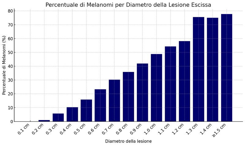
Figure 2.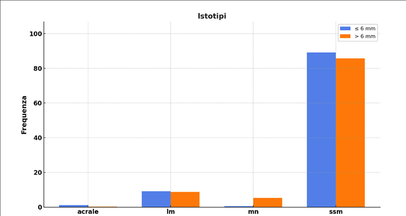
Downloads
Citations


How to Cite

This work is licensed under a Creative Commons Attribution-NonCommercial 4.0 International License.
