

13 | Spatial contexture of tertiary lymphoid structures as a predictor of recurrence in early-stage melanoma
Chiara Bungaro1, Sabino Strippoli1, Daniele Carenza1, Ivana De Risi1, Rossella Villani1, Teresa Squicciarini1, Anna Albano1, Ileana De Roma1, Fabio Mele 2, Margherita Sonnessa2, Concetta Saponaro2, Francesco Alfredo Zito2, Arianna Bozzi3, Benedetta Apollonio1, Raffaella Massafra3, Michele Guida1 | 1Rare Tumors and Melanoma Unit, IRCCS Istituto Tumori "Giovanni Paolo II"; 2Pathology Unit, IRCCS Istituto Tumori "Giovanni Paolo II"; 3Biostatistics and Bioinformatics Unit, IRCCS Istituto Tumori "Giovanni Paolo II", Bari, Italy.
All claims expressed in this article are solely those of the authors and do not necessarily represent those of their affiliated organizations, or those of the publisher, the editors and the reviewers. Any product that may be evaluated in this article or claim that may be made by its manufacturer is not guaranteed or endorsed by the publisher.
Authors
Background: Tertiary Lymphoid Structures (TLS) are organized aggregates of immune cells that form in non-lymphoid tissues in response to chronic inflammation, including cancer. In melanoma, TLS presence within the tumor microenvironment is associated with a pre-existing anti-tumor immunity and linked to improved immunotherapy responses. However, their role in predicting recurrence, particularly in early-stage melanoma, remains unclear.
Methods: We conducted a retrospective analysis of archival primary tumor samples from 65 patients with stage I-II melanoma and negative sentinel lymph node (Table 1). Tumor immune infiltrates and TLS were initially assessed using four-marker immunohistochemistry (IHC) panels targeting CD20, CD4, CD8, and Podoplanin (PDPN), a marker of stromal cells. TLS were objectively identified using the HDBSCAN AI-based clustering algorithm. Tumor samples were further analyzed for high-dimensional phenotyping and spatial analysis using a 17 markers panel with hyperplexed immunofluorescence imaging (HIFI). Associations between TLS features and clinical outcomes were evaluated.
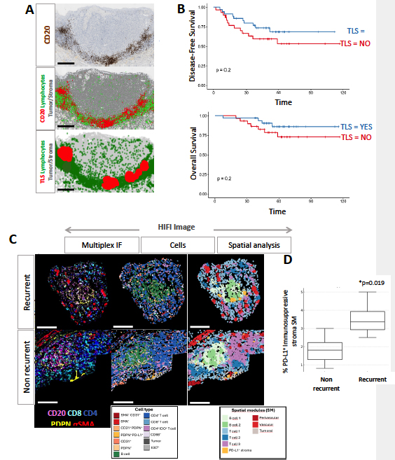
Results: IHC revealed that 88% of patients had peritumoral T cell infiltration, and TLS were detected in 54% of cases. TLS-positive patients had improved disease-free survival (DFS, p=0.2) and overall survival (OS, p=0.2) (Figure 1A, B). To investigate further, HIFI was performed on 8 patients (4 recurrent, 4 non-recurrent), revealing 13 distinct cell types: stromal (n=6), tumor (n=2), and immune (n=5) (Figure 1C). Spatial analysis identified 9 recurrent spatial modules (SM), each representing a distinct pattern of interaction between cell types. One SM, enriched for PD-L1⁺ stromal cells, was significantly associated with recurrence (p=0.019) (Figure 1D).
Conclusions: TLS presence associates with improved DFS and OS in early-stage melanoma. However, beyond their mere presence, TLS spatial organization and local stromal-immune context could influence clinical outcomes. Our data demonstrated that a specific spatial module—defined by the proximity of PD-L1⁺ stromal cells—is associated with disease recurrence.
Table 1. 
All p-values are calculated in relation to recurrence status. Significant associations were identified for Sex (Recurrence less frequent among female patients) and for Ulceration (more common in recurrent cases).
Figure 1A-D.
Downloads
Citations


How to Cite

This work is licensed under a Creative Commons Attribution-NonCommercial 4.0 International License.
